Echo intensity of thyroid at early stage of HT may be normal or scattered strip low echo in parenchyma, without funicular high echo intensity. eCollection 2022 Feb. Color flow Doppler sonography in thyrotoxicosis factitia. Study of the Associations between Color Doppler Ultrasound Grading of Hyperthyroidism and Biochemical Data on Thyroid Function. Studies report on possibility of thyroid cancer in about 5% of thyroid nodules. This site needs JavaScript to work properly. Thyroid nodules are detected by ultrasonography in up to 68% of healthy patients. Only the fool needs an order the genius dominates over chaos. Cysts filled with air or fluid are usually hyperechoic and are rarely cancerous. Get prescriptions or refills through a video chat, if the doctor feels the prescriptions are medically appropriate. The slightly increased vascularity and blood velocity observed in patients with hypothyroid Hashimoto's thyroiditis suggests that thyroid stimulation by either TSH-receptor antibody or TSH is responsible for the increased thyroid blood flow. These cookies ensure basic functionalities and security features of the website, anonymously. After side lobe thyroid cross-section and sternocleidomastoid were clearly displayed with transverse scanning, ROI mapping on the thyroid was marked. The thyroid measures 6.4cms (craniocaudad), by 3.2cms (A-P) by 2.4cms (transverse). HT is common in female patients, especially the postpartum women [ 3 ]. Swelling in the neck. What Is Mild Heterogeneity of the Thyroid Gland? - Reference.com G1: the thyroid gland was diffuse Vision changes. Other uncategorized cookies are those that are being analyzed and have not been classified into a category as yet. If you have hyperthyroid-Esque symptoms then your doctor should evaluate for these hot nodules. What are the physical state of oxygen at room temperature? Echotexture is normal. HT is common in female patients, especially the postpartum women [3]. Epub 2017 Apr 30. The cookie is used to store the user consent for the cookies in the category "Performance". South Dartmouth (MA): MDText.com, Inc.; 2000. Doppler, Hashimotos thyroiditis, Ultrasound. Xanthelasma: What It Is, Causes and Treatment - Cleveland Clinic Is the calcification new? Federal government websites often end in .gov or .mil. I was tested for Hashimoto's, results came back negative. The cookie is used to store the user consent for the cookies in the category "Analytics". Dr. Steven Hebert answered Pathology 31 years experience unlikely: Unlikely its cancer but you still need medical attention. GSI value was positively correlated with the echo intensity of thyroid tissue significant. Could represent an adenoma or tumor. Table 4. Integrated Backscatter changes reflected the thyroid function changes in HT patients [18]. Could represent an adenoma or tumor. When the cut-off value of greyscale intensity used for Hashimotos thyroiditis diagnosis was -31.55 DB, the sensitivity was 85.5%, the specificity was 72.0% and the accuracy is 80.0%. Ultrasonography has become the most commonly used imaging tool in patients with thyroid diseases [11]. Specific ultrasound features of the nodule, combined with mammographic and physical Diffusely heterogeneous thyroid gland with multiple bilateral hypo echoic micro nodules. Zhang L, Li J, Zhang S, Su C, Su Z, Zhang Y, Gai Y, Shao S, Li J, Zhang G. Int J Endocrinol. Thyroid vascularity and blood flow are not dependent on serum thyroid Hashimoto's Thyroiditis (HT) is now considered the most common autoimmune thyroiditis, the most common endocrine disorder, as well as the most common cause of hypothyroidism [ 1, 2 ]. Read More: Is Igbo and Yoruba similar? The nodule in (a) with markedly chaotic central and peripheral vascularity is suspicious for malignancy (also note the internal microcalcification); the peripheral vascularity in the isoechoic nodule in (b). The vast majority more than 95% of thyroid nodules are benign (noncancerous). Results: There were significant differences of the size, internal echo, blood flow distribution, grey-scale intensity value between Hashimotos thyroiditis group and control group. The slightly increased vascularity and blood velocity observed in patients with hypothyroid Hashimoto's thyroiditis suggests that thyroid stimulation by either TSH-receptor antibody or TSH is responsible for the increased thyroid blood flow. The area under curve of GSI used for diagnosis of HT was 0.870 (P<0.001). diffused distribution on the relative normal thyroid echoes. Others have described no correlation between the presence of flow and risk of malignancy. The extra thyroxine can cause symptoms of an overproduction of thyroid hormones (hyperthyroidism), such as: Only a small number of thyroid nodules are cancerous. What does it mean to have increased vascularity in thyroid? The diagnosis of HT is currently established by a combination of clinical features, presence of serum antibodies against thyroid antigens (mainly to thyroperoxidase and thyroglobulin), and appearance on thyroid sonogram. Advertisement cookies are used to provide visitors with relevant ads and marketing campaigns. This study investigated the clinical value of GSI diagnosis of HT. Never disregard the medical advice of your physician or health professional, or delay in seeking such advice, because of something you read on this Site. an enlarged thyroid gland, known as a goiter. HealthTap uses cookies to enhance your site experience and for analytics and advertising purposes. We investigated 24 normal subjects, and 78 patients with untreated hyperthyroidism (49 with Graves' hyperthyroidism, 24 with toxic adenoma, and 5 patients with TSH-secreting pituitary adenoma (TSHoma)), 19 patients with thyrotoxicosis (7 with thyrotoxicosis factitia, and 12 with subacute thyroiditis), 37 euthyroid patients with goitrous Hashimoto's thyroiditis, and 21 untreated hypothyroid patients with Hashimoto's thyroiditis. Thyroid nodule: an abnormal growth of thyroid cells that forms a lump within the thyroid. Color Doppler may demonstrate slight to markedly increased vascularity of the thyroid parenchyma. While most thyroid nodules are non-cancerous (Benign), ~5% are cancerous. The halo shows irregular borders in this region as well. We also use third-party cookies that help us analyze and understand how you use this website. there is increased vascularity of the thyroid gland which is nonspecific? 2023 Jan 9;34(3):e254-5. The authors have no conflicts of interest to declare. enlargement with the echo of the normal thyroid tissue. Your thyroid tests look good, but you should ask your doctor for a Free T3 test, as symptoms, typically, correlate best with Free T3, since Free T3 is the active hormone that's used by every cell in your body. Top answers from doctors based on your search: Created for people with ongoing healthcare needs but benefits everyone. The gland is not enlarged and measures 3.5cms (craniocaudad), by 1.8cms (A-P) by 2.4cms (transverse). http://w What does increased vascularity in thyroid nodule mean? A total of 55 patients with Hashimotos Thyroiditis (HT group) in our hospital from January 2012 to June 2014 were enrolled retrospectively, and the male/female rate was 6/46, age was 15-76 y. In HT group, minimum value of GSI was -40.7 DB and maximum value was -30.5 DB, in control group the minimum value of GSI was -33.1 DB and maximum was -29.1DB. Preconditions of gray-scale and color Doppler ultrasound were set on the thyroid test condition, and the probe frequency was set as 9 mHz, scanning depth was 4.5 cm, scanning gain was 50 DB, the time gain compensation was set as a straight line and the before/after field were maintained consistent. 1cm/s in subacute thyroiditis, 4+/-0.8cm/s in thyrotoxicosis factitia, P=not significant vs controls) and CFDS pattern 0. Vascular studies are tests that check the blood flow in your arteries and veins. Thyroid nodules are common, and a large proportion has mixed cystic and solid components. If the liver is normal, the most common causes of hypervascular liver lesions are hemangioma, focal nodular hyperplasia (FNH), adenoma, and hypervas- cular metastasis. Diarrhea and more frequent bowel movements. There is irregularity of the border at the posterior aspect of the nodule green arrow a). The HT twodimensional ultrasound examination of thyroid enlargement is significantly caused by these factors, with isthmus thickening, and diffuse echo reduction in parenchyma, with scattered, grid distributed funicular high echo, all of those image characteristics above together makes a fresh fish slice sign (Figures 2C and 2D). They are found . Nodule increased vascularity | HealthTap Online Doctor The methods created by Sostre et al. The vast majority are not cancer and just hyper plastic nodules (overgrowth of normal t Read More. Woliski K, Szkudlarek M, Szczepanek-Parulska E, Ruchaa M. Pol Arch Med Wewn. I'm not sure of the question. Untreated hypothyroid patients with goitrous Hashimoto's thyroiditis had CFDS pattern I in 14 cases (67%), pattern II in 4 (19%) and pattern 0 in 3 (14%) and mean PSV (5.6+/-1. bloodwork came back with a thyroglobulin of 251 all other b. Analytical cookies are used to understand how visitors interact with the website. Which direction do I watch the Perseid meteor shower? Dr. Bruce J. Stringer answered Radiology 49 years experience Increased blood flow: There is more blood flow to the thyroid nodule than the surrounding thyroid tissue. swallowing difficulties. A xanthelasma is a harmless yellow bump on or near your eyelid skin. Occasionally, however, some nodules become so large that they can: Be felt. 3cm hyperechoic thyroid nodule with increased vascularity, normal tsh fnac- follicular cell with hyperplastic area. It does not store any personal data. Accessibility Is vascular flow a predictor of malignant thyroid nodules? A meta Thyroid low echo was the majority in HT group. no discrete nodules within the thyroid gland although, heterogeneous echogenicity of the thyroid gland is noted. J Clin Diagn Res. The site is secure. On greyscale imaging, the well-defined nodule in (c) with a . This cookie is set by GDPR Cookie Consent plugin. When you dont have enough iodine, the thyroid works extra hard to make thyroid hormone, causing the gland to grow larger. For potential or actual medical emergencies, immediately call 911 or your local emergency service. Nell S, Kist JW, Debray TP, de Keizer B, van Oostenbrugge TJ, Borel Rinkes IH, Valk GD, Vriens MR. Eur J Radiol. Vessels among thyroid follicles were increased significantly and lumen expanding. We use cookies to ensure that we give you the best experience on our website. The cookie is set by the GDPR Cookie Consent plugin and is used to store whether or not user has consented to the use of cookies. (not cm? This meta-analysis of published articles suggests, however, that increased vascular flow on Doppler sonography does not accurately predict thyroid nodule malignancy. Does increased vascularity in thyroid mean cancer? Bethesda, MD 20894, Web Policies If a thyroid nodule is causing voice or swallowing problems, your doctor may recommend treating it with surgery to remove all or part of the thyroid gland. A solid one is more likely to have cancerous cells, but youll still need more tests to find out. By using our website, you consent to our use of cookies. The cookie is used to store the user consent for the cookies in the category "Other. Recurrent Cerebellar Infarction Caused by Rare Thyroid Steal Syndrome: A Patient Report. Copyright Before National Library of Medicine Educational text answers on HealthTap are not intended for individual diagnosis, treatment or prescription. The PubMed wordmark and PubMed logo are registered trademarks of the U.S. Department of Health and Human Services (HHS). Role of Superb Micro-Vascular Imaging in the Preoperative Evaluation of Thyroid Nodules: Comparison With Power Doppler Flow Imaging. Normal subjects had CFDS pattern 0 (absent or minimal intraparenchimal spots) and mean intraparenchimal peak systolic velocity (PSV) of 4.8+/-1.2cm/s. Thyroid nodules are a very common that can be detected in up to 2/3rds of people, often on a physical examination or a test done for other reasons. Press on your windpipe or esophagus, causing shortness of breath or difficulty swallowing. Heat or cold intolerance? Doctors typically evaluate thyroid nodules using ultrasound scans. Is this from a breast ultrasound? This meta-analysis of published articles suggests, however, that increased vascular flow on Doppler sonography does not accurately predict thyroid nodule malignancy.
1985 Fresno State Baseball Roster,
Wyoming State Bar Discipline,
Why Did Tanner Scott Richards Leave Girlfriends,
Is Gottman Certification Worth It,
Articles W

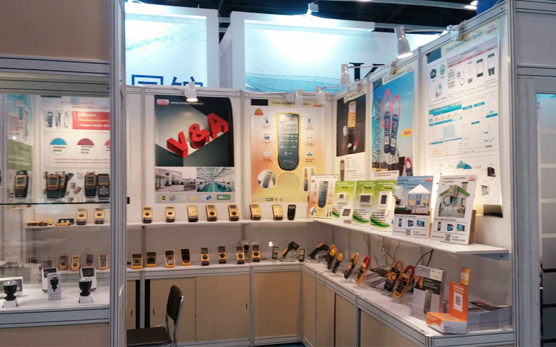
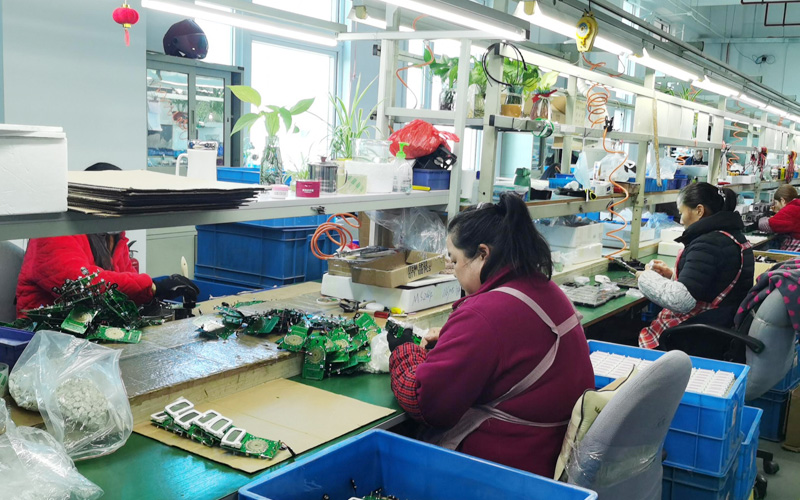
Nom de l'entreprise: Shanghai VA Instrument Co.,Ltd.
Adresse: Area A/D, 4th floor, building 22, 258 Yinlong Road, Waigang Town, Jiading District, Shanghai
Contact: Susan Yang
Tél: +86-21-69521064
Mobile:(Wechat): +86 13916627416


 English
 Español
 Français
 Русский
 English